Ionizing Radiation and Safety
Branch note: This page deepens one part of Nuclear Physics.
Official 9749 focus
Outcome 20(t) requires a qualitative discussion of both direct and indirect effects of ionising radiation on living tissues and cells. Radiation properties and background are also core Topic 20 knowledge. Contamination/irradiation, time–distance–shielding, detectors, applications and waste are useful context but not additional explicit recall outcomes.
Overview
Ionizing Radiation and Safety covers the hazards, uses, detection, and safe handling of ionizing radiation.
Ionizing radiation has enough energy to remove electrons from atoms, producing ions. This can be useful in medicine and industry, but harmful to living tissue.
It is the applied branch in the nuclear-physics block. It explains:
- what ionizing radiation is
- types of radioactive emissions
- biological hazards of radiation
- contamination and irradiation
- radiation protection methods
- radiation detection
- useful applications in medicine and industry
This topic connects nuclear-physics ideas to real-world safety and technology.
This topic links closely with:
Core Ideas
- ionizing radiation can remove electrons from atoms and molecules, producing ions
- alpha, beta, and gamma radiations have different ionising and penetrating powers
- radiation may damage a critical molecule directly or act indirectly through reactive species produced by ionising water or other molecules
- cellular outcomes include repair, mutation, loss of function or cell death; tissue effects and cancer risk depend on dose and context
- background radiation has natural and human-made origins and affects detector readings
Useful enrichment/application context: hazard also depends on exposure route, contamination differs from irradiation, and risk can be reduced using time, distance and shielding.
What Is Ionizing Radiation?
Ionizing radiation is radiation that can knock electrons out of atoms or molecules.
This causes:
- ion formation
- chemical changes
- biological damage in cells
Common nuclear ionizing radiations:
- particles
- particles
- rays
The safety question is not just “which radiation is strongest?” The hazard depends on how the radiation interacts with matter, whether the source is inside or outside the body, and how long the exposure lasts.
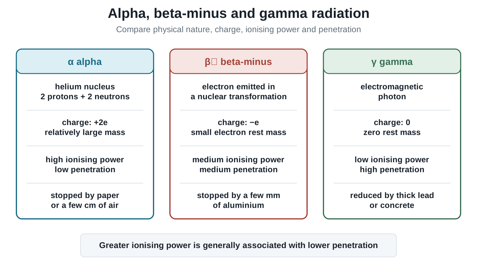
Figure: Compare alpha, beta-minus and gamma radiations by physical nature, charge, relative ionising power and penetration. The comparisons are qualitative trends under comparable conditions, not fixed numerical values. Gamma radiation is highly penetrating, so shielding reduces or attenuates it rather than guaranteeing complete stopping in one thin layer.
Common Types of Ionizing Radiation
Alpha Radiation ()
- helium nucleus,
- charge =
- relatively large mass compared with beta particles
- highly ionising
- weakly penetrating
Beta Radiation ()
- in the core example, a fast electron created and emitted during a beta-minus nuclear transformation
- charge
- moderately ionising
- moderately penetrating
Enrichment
Positron emission is not required by 9749. If encountered in supplied context, a positron has charge , but this is not core recall.
Gamma Radiation ()
- electromagnetic radiation
- no charge
- zero rest mass
- weakly ionising
- highly penetrating
Comparison Table
| Radiation | Nature | Charge | Relative Mass | Ionising Power | Penetrating Power | Typical Shielding |
|---|---|---|---|---|---|---|
| helium nucleus, | relatively large | high | low | paper, air, or outer dead skin layer | ||
| electron | small | medium | medium | a few millimetres of aluminium | ||
| electromagnetic radiation | zero rest mass | low | high | thick lead or concrete |
Key Reminder
Ionising power and penetrating power are different properties.
Sources of Background Radiation
Background radiation is the radiation detected when the particular source being investigated is absent. It originates from the surroundings and must be allowed for when a source count rate is measured. Its count fluctuates randomly, so a mean background rate is estimated over a suitably long interval.
Main sources include:
Natural Sources
- cosmic rays from space
- radioactive rocks and soil
- radon gas in air
- naturally radioactive food and water
- radiation from living organisms
Artificial Sources
- nearby medical, industrial or research sources, depending on the measurement setting
- residual fallout and nuclear-industry sources where relevant
Biological Effects
Ionising radiation transfers energy to atoms and molecules in tissue, forming ions or excited molecules and sometimes breaking chemical bonds. Damage can arise through two distinct causal routes.
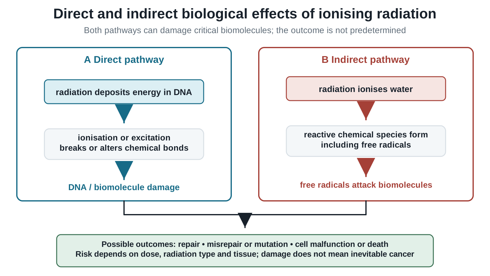
Figure: In the direct route, radiation deposits energy in a critical biological molecule such as DNA, causing ionisation, excitation or bond damage. In the indirect route, radiation first ionises water or another molecule, producing reactive chemical species such as free radicals that subsequently damage DNA or other cell structures. Either route may be repaired or may lead to malfunction, mutation or cell death; the outcome is probabilistic, not inevitable.
Direct Effect
Radiation deposits energy in a critical biological molecule directly. For example, direct ionisation or excitation of DNA may break a molecular bond or strand, alter genetic information, or interfere with replication and normal cell control.
Indirect Effect
Radiation ionises water or another molecule in the cell, creating reactive ions or free radicals. These species can then chemically attack DNA, proteins or membranes. Radiation therefore need not strike DNA directly to cause damage.
From Molecular Damage to Biological Outcome
Cells can repair some damage. If repair is unsuccessful or incorrect, possible outcomes include loss of cell function, cell death, mutation, uncontrolled cell division, increased cancer risk and tissue damage when many cells are affected.
These outcomes are probabilistic, not guaranteed after every exposure. Likelihood and severity depend on absorbed energy, radiation type, exposed tissue, exposure route and other biological factors. Activity alone is not a complete measure of biological risk.
Ionisation in Tissue
Radiation ionises atoms in cells, disrupting chemical processes.
Cell Damage
This may cause:
- cell death
- tissue damage
- burns at high dose
Mutation and Cancer Risk
DNA damage may lead to:
- mutations
- cancer
- hereditary effects in some cases
Risk generally increases with dose, but this describes probability; it does not mean every irradiated cell becomes cancerous.
Enrichment: Contamination vs Irradiation
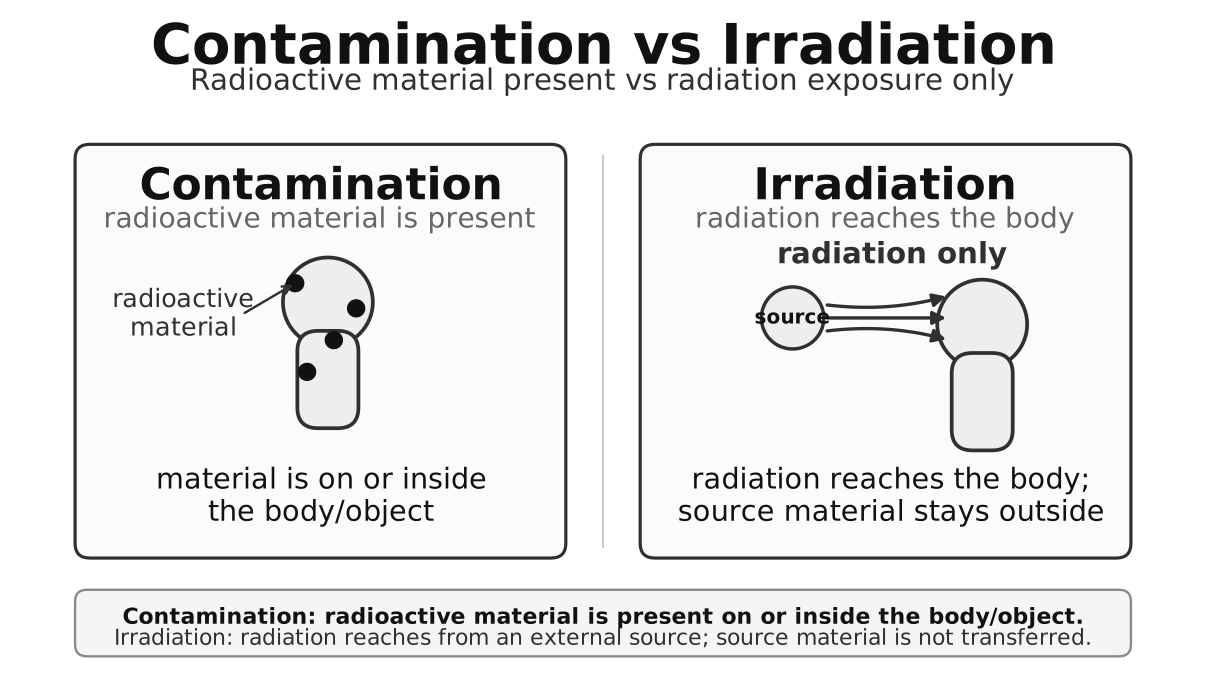
Figure: Contamination means radioactive material is on or inside the body, whereas irradiation means exposure to radiation from an external source.
Contamination
Radioactive material is deposited on or inside an object or person.
Examples:
- radioactive dust on skin
- inhaled radioactive particles
Hazard continues while the source remains present.
Irradiation
An object or person is exposed to radiation from an external source.
When the source is removed, the exposure stops.
Important Difference
A person can be irradiated without becoming radioactive.
Enrichment: Internal vs External Hazard
External Exposure
The radiation source is outside the body.
- is often serious because it penetrates deeply
- is often less serious externally
Internal Exposure
The radioactive source is inside the body by:
- inhalation
- swallowing
- wound entry
Then:
- can be very dangerous because of strong ionisation in nearby tissue
Enrichment: Factors Affecting Hazard
1. Type of Radiation
- can be especially dangerous inside the body
- is often more dangerous externally because of high penetration
2. Activity
Higher activity means more decays per second.
3. Exposure Time
Longer exposure time increases dose.
4. Distance
Greater distance usually reduces exposure.
5. Shielding
Suitable materials reduce exposure.
6. Internal vs External Exposure
Internal sources can greatly increase hazard, especially for emitters.
Enrichment: Safety Principles
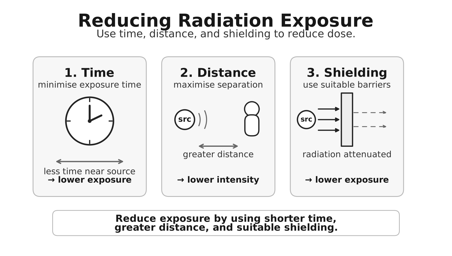
Figure: Radiation exposure is reduced by minimizing time, maximizing distance, and using suitable shielding.
Minimise Time
Spend less time near the source.
Maximise Distance
Increase separation from the source.
Use Shielding
Examples:
- paper or the outer dead layer of skin for
- aluminium for
- lead or concrete for
Additional Practices
- remote handling tools
- sealed containers
- protective clothing
- warning signs
- monitoring badges
Enrichment: Uses of Ionizing Radiation
Medicine
- cancer radiotherapy
- medical imaging
- sterilising equipment
Sterilisation
Ionizing radiation kills microorganisms in food or medical tools.
Tracers
Radioactive tracers track movement in:
- medicine
- pipelines
- biological systems
Industrial Inspection
Ionizing radiation is used to detect:
- cracks
- thickness changes
- leaks
Agriculture and Food
- sterilisation
- food preservation
Research
- radioactive tracers
- detectors
Enrichment: Waste and Disposal Context
Radioactive waste may remain hazardous for long periods.
Management may require:
- shielding
- secure storage
- controlled transport
- waiting for decay where appropriate
Waste from fission reactors can be significant.
See Nuclear Fission.
Enrichment: Radiation Detection
Radiation usually cannot be sensed directly.
Common detectors include:
Geiger-Muller Tube
Detects ionising events and gives count rate.
Film Badge or Dosimeter
Measures worker exposure over time.
Cloud Chamber
Shows visible particle tracks.
Count Rate Correction
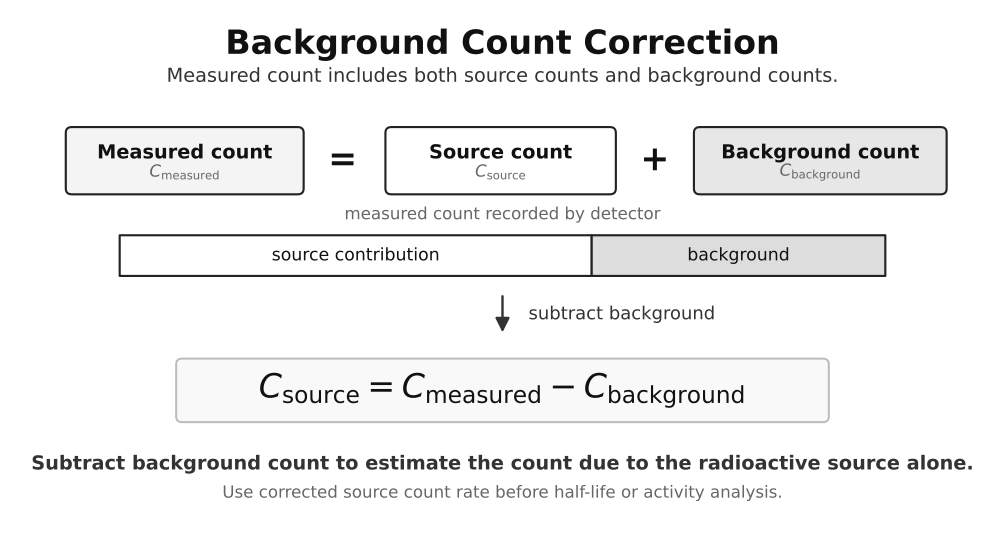
Figure: Source count rate is measured count rate minus background count rate.
Measured count rate includes background. Subtract the mean background rate measured with the source absent:
For fuller detector and monitoring detail, see Radiation Detection and Monitoring.
Short Worked Examples
Example 1: Best Shield for Gamma Radiation
Question: Which material is most suitable?
Answer:
Dense thick materials such as lead or concrete.
Example 2: Alpha Source Outside the Body
Answer:
It is usually less dangerous externally because alpha particles are stopped by paper, air, or the outer dead layer of skin.
Example 3: Why an Internal Alpha Source Is Dangerous
Answer:
Alpha particles strongly ionise nearby tissue inside the body.
Example 4: How To Reduce Exposure Quickly
Answer:
Reduce exposure time, increase distance, and use appropriate shielding.
Formula Reminder
Exam Relevance
Assessed Topic 20 core
Students should be able to:
- compare alpha, beta, and gamma hazards qualitatively
- explain both direct damage to critical molecules and indirect damage through reactive species formed after water ionisation
- connect molecular damage qualitatively to mutation, cell death, cancer risk and tissue effects
- identify origins and significance of background radiation
Useful enrichment and application context
Students may also use this note to:
- distinguish contamination from irradiation
- choose appropriate shielding
- explain why internal and external hazards can differ
- describe basic safety principles and applications
Common Exam Traps Overview
Students often confuse:
- contamination with irradiation
- ionising power with penetrating power
- assuming alpha is always least dangerous
- forgetting background radiation can be natural
- choosing the wrong shielding
- assuming more penetrating always means more biologically damaging
- naming cancer or mutation without explaining either the direct or indirect ionisation pathway
- assuming that every exposure produces a deterministic biological outcome
See Ionizing Radiation and Safety Common Exam Traps.
Quick Revision Summary
- ionizing radiation removes electrons from atoms
- , , and are common nuclear examples
- radiation can damage cells and DNA
- direct damage acts on a critical molecule itself; indirect damage acts through reactive species formed elsewhere, commonly after water ionisation
- contamination means radioactive material is present
- irradiation means exposure from a source
- reduce risk using time, distance, and shielding
- radiation has many medical and industrial uses
Final Memory Line
Radiation can be highly useful or highly harmful. Safety depends on understanding ionisation, exposure, and protection.